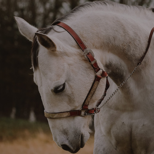
Colic
Colic in horses – what is it?
In strict definition, colic is a term that describes a painful problem in the horse’s abdomen. In general, the source of abdominal pain can originate from any location within the horse’s abdomen but most commonly, colic occurs if parts of the horse's gut becoming impacted or from getting in an unphysiological placement within the abdomen (blood supply can be shut off) as well as intestinal spasms (gut wall overstretching by gas or feed material). In significantly rarer cases, abdominal pain can also arise from the genitourinary tract (bladder or ureteral stones, ovarian tumors, uterine torsion) or be due to the presence of tumors in the abdominal cavity (false colic).
Colic is a frequent internal emergency and unfortunately still one of the most common causes of mortality in horses although the chances for surviving are far better than ten years ago.
Colic of digestive origin
The digestive tract in horses is about 30 meters long and is made up of stomach, small intestine, small colon, caecum, large colon, and rectum. Due to this specific anatomy of the horses’ gastrointestinal tract as well as the high sensitivity towards stress (mainly due to several changes in feed, environment, or weather) and pain, horses are more prone to suffer from colic than most other mammalian species.
Gastric ulcers
Gastric ulcers are a wide-spread and common issue not only in race- and sports horses but more and more also occurring in leisure horses. Beside changes in feeding habits (diets high in starch and low in forage) and unbalanced housing conditions (single stall living, limited access to pasture, intensive exercise) also frequent treatment with NSAIDs can elicit the occurrence of gastric ulcers. Horses with gastric ulcers are often suffering from abdominal pain most frequently after feeding and can develop signs of in colic in severe cases of ulceration.
Ileus
In the case of an ileus (Entrapment in existing abdominal structures, lipoma pendulans), the feed bolus from the stomach and the intestine backs up in front of the obstruction. In addition, fluid from the bloodstream continuously enters the small intestine, which also leads to a buildup at the obstruction site. The intestinal wall is severely stretched prior to the occlusion, and feed, fluid, and resulting gases back up into the stomach, which is also stretched (secondary gastric overload). Beside the risk of gastric rupture, the loss of fluid from the bloodstream into the intestine can lead to a shock as less and less fluid is available to the horse's bloodstream.
The small intestine is suspended in the horse's abdomen from the mesentery. If the intestine is heavily filled, this can lead to a twist around its own mesentery which causes the intestine and mesentery to become knotted which marks an emergency which requires surgery immediately.
Intestinal causes of colic
Due to a specificity in the horses’ gastrointestinal anatomy, the large intestine (colon) is predominantly “floating” in the abdomen and only fixed at few points in the abdominal cavity. Thus, the colon can easily be displaced and is predestinated for bottlenecks at some places which can provoke obstruction.
Spasmic (gas) colic
Abrupt changes in food habits (too much gras, high intake of grain) due to microbiome hyper fermentation are the main reason for this type of colic. If a treatment is necessary, it mostly consist in giving NSAIDs or opioids to control pain, and spasmolytic drugs to alleviate painful contractions of the intestine.
Obstruction colic
Overconsumption of forage, insufficient water intake or lack of movement can provoke this kind of colic which often occurs in winter (low intensity to drink, high demand for forage). Medical treatment with NSAIDs and spasmolytic substances is often sufficient in addition to intake of laxatives by nasogastric intubation. Intravenous rehydration with sterile liquids can become necessary in some cases.
Dislocation of the colon
It is assumed that increased intestinal movement, strong filling of intestinal segments and strong gas development in the intestine contribute to the fact that intestinal parts are displaced. Medical treatment with NSAIDs and spasmolytic substances is sometimes not sufficient, and surgery might be necessary replace the colon in its physiological position.
Colon torsion
The large intestine rotation basically arises from a preceding large intestine displacement. The formation of gas by the microbiota in the colon plays a major role here as it causes a natural flotation in a part of the colon, making it lighter and allowing it to move upward. Due to a complex process, in such cases the colon can rotate around itself which cuts off the blood supply. This form of colic is one of the most dangerous and painful and if the blood supply is not restored by untwisting the rotation, the colon dies which is lethal to the horse as well. Therefore, horses affected by colon torsions always require surgery, where the colon is untwisted again, and the contents of the colon are emptied.
Intussusceptions
A part of the intestine slides into the intestine in front of it like a telescope. Intussusception of the cecum is described to be the most common cause of cecal obstruction especially in young horses (< 3 years). Intestinal parasites and other agents that are altering the motility of the gastrointestinal tract have been implicated as potential risk factors. Colic signs vary from severe acute abdominal pain requiring immediate surgical intervention to intermittent, chronic signs of colic.
Colitis X
The cause is an inflammation which most commonly occurs in horses post-colic-surgery. Stress, antibiotic treatment, surgical interventions, worm infestation, food deprivation are suspicious to trigger colitis-X however the detailed pathology is still unclear. It is assumed that a change in the composition of the intestinal flora is of decisive importance as certain strains of clostridia (Clostridium difficile and Clostridium perfringens) proliferate disproportionately in the intestine of sick horses, releasing endotoxins which are absorbed through the intestinal wall into the blood, leading to an endotoxin shock. In addition, the intestinal wall is inflamed which leads to increased perfusion due to which a lot of fluid is lost from the blood vessels into the intestine (Risk of volume deficiency shock). The severe inflammation of the intestinal wall can lead to the death of parts of the intestinal wall. Colitis-X must be treated intensively as early as possible to increase the chances of surviving for the horse. Anti-inflammatory drugs, intestinal stimulation drugs, intravenous liquid infusions (electrolytes), easily digestible diet are the first-choice method.
Symptoms of colic in horses
Various clinical signs are associated with colic and the clinical signs mainly depend on the cause of the colic as well as the personality of each horse.
Mild cases:
Restlessness, flank wa, pain face (lip curling, ears set backwards), pawing the ground, poor appetite, change in drinking behavior, stretching outtching / kicking
Moderate cases:
Posturing to urinate frequently, Straining to defecate, lying down and getting back up or lying on their side for long periods, depression, changed number of bowel movements
Severe cases:
Violent rolling in uncontrolled and dangerous manner, moderate to heavy sweating, rapid breathing, increased heart rate, decreased number of bowel movements,
What to do in case of colic?
Colic is a potentially life-threatening emergency in horses. If a horse shows moderate to severe colic symptoms, urgent veterinary examination is a must. Horses with mild colic symptoms should be walked around (only walk, no trot or canter) for around 10-15 minutes if it makes them feel better.
CAUTION: Pleuritis, tying up or laminitis can result in similar signs like colic. Horses suspicious to suffer from such a disease should NOT walked as this can lead to further pain.
If the symptoms persist or getting severe, the vet also should be called for further examination immediately. The horse’s feed should also be removed from the stable while fresh water needs to be provided at any time.
Before the vet is arriving in the stable, you can support by providing the vital parameters (heart rate/minute, body temperature, breathing rate/minute) as well as the capillary refill time already taken from your horse.
Veterinary examination
To determine the possible reason for colic in the horse, the vet is going to ask several questions and will perform a physical exam of the horse.
The questions may include one or more of the following:
Changes in feed, diets high in grain (low in forage), long periods between forage / feeding, recent travels (stress), previous episodes of colic, deworming status, medication, access to toxins (herbs, fungal etc.), Sand ingestion, last dental examination, gastric ulcers occurring, decrease / increase in water intake, Changes in routine (exercise)
The physical examination should include:
Heart rate, respiratory rate, rectal temperature, auscultation of presence or absence of gastrointestinal sounds, abnormal color of mucous membranes, capillary refill time, skin turgor, digital pulses of the hooves, abdominal distention, rectal examination
Depending on the results, the vet may choose to perform one or all of the following further examinations: Blood examination, passing of a nasogastric tube into the stomach (accumulation of fluid in the stomach), obtaining a sample of the fluid that surrounds the intestines from the abdomen (abdominocentesis), abdominal ultrasound, gastroscopy, x-ray
Risk factors – How to prevent colic?
Horses are animals of habit and although every change in their lifestyle, environment, activity, or feed is a risk factor for colic that can be managed, there are some types of colic that aren’t preventable.
Changes in diet
Prevention: Feed changes always should take place slowly (for 1-2 weeks starting with mixing one fourth of the new with 3 parts of the current feed) and not during variable weather periods. Feed grain and any type of pelleted feed only when necessary.
High parasite infestation
Routine parasite prevention: Fecal egg count 2-4 times per year (or as recommended by your vet) along with a routine deworming program within the barn.
Sand ingestion
Prevention : Avoid feeding on the ground in sandy areas and always provide your horse with a balanced mineral feed (e.g., MEGA BASE) to avoid mineral deficiencies that horses sometimes try to compensate via eating sand.
Dental issues
Prevention: Routine teeth control and examination of the oral cavity at least once per year ensures properly and thoroughly chewing of the feed and assures sufficient supply of saliva as a buffer substance for the stomach.
Decrease in water intake
Prevention : Horses prefer to drink out of buckets which is likely due to their ability to drink large amounts more quickly.
In the winter, the water provided need to be prevented from freezing (floating objects, warm water). Additionally, horses will drink more in winter if the water is warm.
High grain diets
Prevention : Increase forage ratio (which should be at least two third of the horse’s entire daily diet) by providing hay several times per day. Access to pasture should be granted for more than 4 hours per day. Chose also different sources of forage to increase the intake of fiber (straw, hay cobs).
Common use of NSAIDs
Prevention : Try to reduce the use of NSAIDs to the lowest level possible as some substances (e.g., Phenylbutazone, Flunixin Meglumine) can increase the risk for colic (ulceration) or at least hide early symptoms. Always discuss the correct dose with your vet and avoid using large amounts or long-term use.
Weather changes
Prevention : Many horses have an altered thirst behavior during abrupt weather changes and are prone to obstruction colic. Especially in winter, horses should have sufficient opportunities for water intake. In addition, only limited pasture access should be granted during the transitional period, as the fructan content in the grass can fluctuate massively with strong temperature fluctuations in spring and fall and thus also increase the risk of colic (and laminitis).
Changes in routine or exercise
Prevention : Make only gradual changes in housing and exercise (as well as in diet) if necessary. Reduced exercise does not necessarily lead to colic but is associated with a higher risk. Horses who are injured or having a break from exercise should not be bedded on straw as they may eat their bedding which can lead to overweight and increased risk for colic as well.
Always strictly monitor any horses that have displayed any signs of colic previously!
Dr. med. vet. Julie Dauvillier
Dr. med. vet. Caroline Fritz
